Ancient Roman Sculptures as Early Depictions of Occult Spinal Dysraphism
– 6 min read


Occult Spinal Dysraphism and Its Cutaneous Stigmata in Clinical Neurosurgery
This comprehensive study takes readers on an extraordinary journey from modern neurosurgical clinics to the marble halls of ancient Roman museums, examining how subtle cutaneous markers of spinal anomalies may have been preserved in stone for nearly two millennia. The research presents a fascinating intersection of contemporary medical knowledge and archaeological investigation, suggesting that ancient sculptors may have unknowingly documented congenital spinal conditions with remarkable accuracy.
Contemporary neurosurgeons recognize that specific dermatological features on the lower back can indicate hidden spinal cord abnormalities. These cutaneous stigmata include localized patches of hair, small dimples or pits, subtle asymmetries, and minor skin depressions along the spinal midline. Collectively termed occult spinal dysraphism, these conditions represent developmental anomalies where the spine and spinal cord failed to form completely normally during embryogenesis, yet remain concealed beneath intact skin.
The term "occult" simply denotes "hidden" - these spinal defects are not immediately obvious upon casual observation but may be revealed through careful examination of surface markers. Modern diagnostic approaches utilize ultrasonography and magnetic resonance imaging to investigate these subtle external signs, often leading to surgical intervention when underlying tethered cord syndrome, split cord malformations, or dermal sinus tracts are discovered.
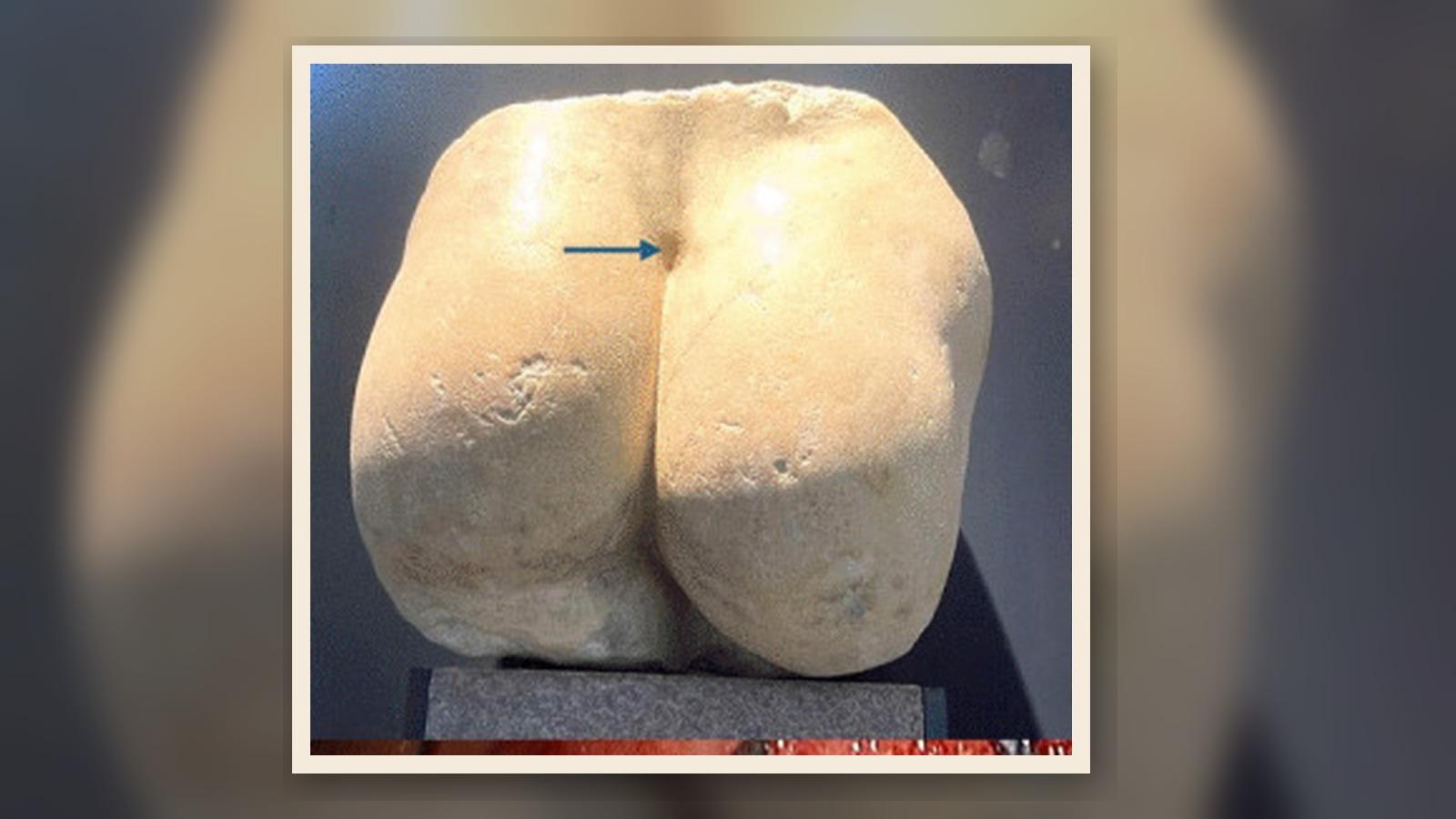
The first compelling example emerges from Cremona, a northern Italian city renowned for violin craftsmanship but equally significant for its Roman heritage. During archaeological excavations near the Palazzo Comunale, researchers discovered a white marble torso fragment dating to the first and second centuries AD. This sculpture, now housed in the San Lorenzo Archaeological Museum, depicts only the posterior aspect of a human form, extending from shoulders to the superior buttock region.
Upon detailed examination, the torso reveals a precisely carved oval depression in the lower lumbar region, positioned directly above the intergluteal cleft. This feature measures approximately four by five centimeters, with remarkably smooth edges and a shallow but deliberate excavation. The surrounding marble surface shows intentional working around this anomaly, while subtle asymmetry between the left and right gluteal regions suggests the sculptor was faithfully reproducing an actual anatomical variation.
The morphology and location of this carved depression correspond precisely to what contemporary physicians recognize as a dermal sinus tract. In living patients, such midline openings present as small, often pinpoint apertures that may extend internally toward the spinal canal through narrow epithelium-lined tunnels. These congenital anomalies pose significant clinical concerns as potential pathways for ascending infection or indicators of tethered cord syndrome.
When neurosurgeons encounter similar presentations in pediatric patients today, imaging studies typically follow to delineate the tract's depth and relationship to neural structures. Surgical exploration often reveals connections to the dura mater or spinal cord itself, necessitating careful dissection to prevent neurological complications. The Cremona statue appears to document this exact anatomical variant with extraordinary fidelity, suggesting Roman sculptors possessed keen observational skills and commitment to realistic representation.
The second remarkable specimen resides within the Vatican's Museo Pio-Clementino, where a Roman marble copy of a fourth-century BC Greek original depicts a satyr supporting the infant Dionysus. While the frontal aspect presents familiar Hellenistic artistic elements, the posterior view reveals an unexpected detail that bridges ancient mythology with modern neurosurgical knowledge.
Carved along the satyr's dorsal spine runs a narrow, vertical tuft of hair, precisely positioned in the mid-to-upper lumbar region. This feature differs markedly from generalized body hair texturing found elsewhere on mythological figures. Instead, it presents as a focused, elevated patch following the spinal midline with remarkable anatomical accuracy.
Contemporary medical literature describes this exact presentation as a "faun's tail" - ironically employing mythological terminology to describe a pathological finding. Focal midline hirsutism in the lumbar region frequently overlies split cord malformations, where the spinal cord divides into two strands over several vertebral segments. Associated conditions may include tethered cord syndrome, intraspinal lipomas, or diastematomyelia.
The Vatican satyr presents an intriguing diagnostic puzzle. While satyrs traditionally exhibit excessive hair as markers of their hybrid nature, the specific location, morphology, and isolated character of this carved tuft mirrors clinical presentations documented in modern pediatric neurosurgery. The precision suggests the sculptor may have observed this feature on a human model, inadvertently preserving evidence of an underlying spinal anomaly in mythological form.
These Roman examples form part of a broader archaeological pattern spanning continents and millennia, where ancient artists documented unusual anatomical variants with striking accuracy. Egyptian temple reliefs frequently depicted individuals with achondroplastic dwarfism, showing characteristic shortened limbs and enlarged cranial features. These figures often held prominent religious or ceremonial roles, suggesting social acceptance rather than marginalization.
Pre-Columbian American cultures produced ceramic works showing diverse congenital conditions. Moche pottery from Peru displays figures with what modern physicians would recognize as lateral facial clefts, while Maya ceramics document cranial dysmorphism consistent with premature suture fusion syndromes. Mesoamerican terracotta figurines show infants with dorsal masses suggestive of open spinal dysraphism, providing three-dimensional documentation of myelomeningoceles centuries before medical terminology existed.
Particularly relevant to spinal anomalies are pre-Hispanic figurines depicting individuals with fused lower limbs, possibly representing sirenomelia or caudal regression syndrome. These artistic works may have influenced mermaid mythologies while simultaneously preserving medical observations. The consistent accuracy across cultures suggests ancient artists served as unwitting anatomical documentarians, recording congenital variations with remarkable clinical precision.
The findspot contexts of these artifacts reveal important social dimensions. Many originated from domestic settings, religious shrines, and burial assemblages rather than medical contexts. Their presence among grave goods and ritual deposits indicates that individuals with visible congenital differences maintained significant cultural roles as priests, entertainers, divine intermediaries, or protected community members.
The Cremona torso's discovery near civic buildings suggests public display or commemoration of local citizens, including those with anatomical variants. Similarly, the Vatican satyr's mythological context may reflect artistic traditions that incorporated observed human diversity into divine narratives, creating inclusive spiritual iconography that celebrated rather than concealed physical differences.
Contemporary understanding of embryological development provides crucial interpretive frameworks for these archaeological findings. Neural tube formation occurs during the third and fourth weeks of gestation, when the presumptive brain and spinal cord separate from surface ectoderm. Incomplete separation may result in persistent connections between skin and neural structures, manifesting as dermal sinus tracts, focal hirsutism, or cutaneous dimpling.
Split cord malformations arise when the developing spinal cord fails to fuse properly, creating dual neural tubes over variable segments. Associated cutaneous markers include midline hair tufts, skin tags, or pigmented patches that serve as surface indicators of deeper anomalies. Tethered cord syndrome results when these abnormal connections restrict spinal cord mobility during growth, potentially causing progressive neurological dysfunction.
Modern imaging techniques reveal the complex three-dimensional anatomy underlying these subtle surface signs. Ultrasonography provides initial screening for spinal canal connections, while magnetic resonance imaging delineates precise relationships between cutaneous markers and neural structures. Surgical management typically involves microsurgical techniques to disconnect abnormal tethering while preserving neurological function.
This investigation demonstrates the value of applying contemporary medical knowledge to archaeological interpretation, creating new dialogues between clinical practice and historical research. The precision with which ancient sculptors documented anatomical variants suggests systematic observation and artistic commitment to realistic representation that transcended purely aesthetic considerations.
The Cremona torso and Vatican satyr become more than decorative objects when viewed through modern medical perspectives. They represent potential case studies preserved in stone, offering insights into the prevalence and social acceptance of congenital conditions in antiquity. Their museum contexts provide accessible laboratories for continued interdisciplinary investigation.
Future research directions might include systematic surveys of museum collections using clinical criteria to identify additional examples of documented congenital conditions. Digital imaging techniques could enhance surface detail analysis, while collaborative networks between medical professionals and archaeologists could refine diagnostic criteria for artistic representations.
The broader implications extend beyond individual case identification to encompass questions about ancient medical knowledge, social attitudes toward physical difference, and the role of artists as inadvertent medical recorders. These marble fragments from Cremona and Rome may represent humanity's earliest preserved "images" of spinal anomalies, predating modern radiological documentation by nearly two thousand years.
Through careful reexamination of familiar archaeological materials with contemporary medical insight, this study opens new avenues for understanding both ancient artistic practices and the deep historical continuity of human anatomical variation. The silent testimony of these carved backs speaks across millennia, connecting modern surgical suites with ancient workshops through the enduring language of careful observation and faithful representation.
Original source article
https://doi.org/10.1007/s00381-026-07258-0


Comments